双语学习分享 学习笔记:肺炎链球菌肺炎
- 2026-11-16 15:29:31
双语学习分享 学习笔记:肺炎链球菌肺炎肺炎链球菌肺炎是社区获得性肺炎(CAP)最常见类型,由肺炎链球菌感染所致。随着抗生素广泛应用,典型临床表现越来越少,临床识别时需要熟悉它的经典特点、不典型变化、诊疗要点,避免漏诊、误诊。 Pneumococcal pneumonia is the most common type of community-acquired pneumonia (CAP), caused by Streptococcus pneumoniae. With the widespread use of antibiotics, classic manifestations are becoming less common. Clinicians need to be familiar with its typical features, atypical variations and key points of diagnosis and treatment to avoid missed diagnosis and misdiagnosis. 肺炎链球菌(SP)是革兰阳性球菌,有荚膜,荚膜多糖是主要毒力因子,一共分为86个血清型,1、3、4、19、23型致病力较强。该菌是上呼吸道常驻定植菌,当机体免疫力下降时,有毒菌株侵入肺部引发感染。细菌不产生毒素,不造成组织坏死空洞;致病核心是荚膜多糖诱发肺泡炎症渗出,病变从肺外周开始,容易累及胸膜,形成渗出性胸膜炎。 Streptococcus pneumoniae (SP) is a Gram-positive coccus with a capsule. Capsular polysaccharide is its major virulence factor, and there are 86 serotypes in total. Serotypes 1, 3, 4, 19 and 23 have strong pathogenicity. This bacterium normally colonizes the upper respiratory tract. When host immunity declines, virulent strains invade the lung and cause infection. SP does not produce toxins and will not cause tissue necrosis or cavity formation. Its pathogenic mechanism mainly relies on capsular polysaccharide to trigger inflammatory exudation in alveoli. Lesions start from the periphery of the lung and easily involve the pleura, leading to exudative pleurisy. 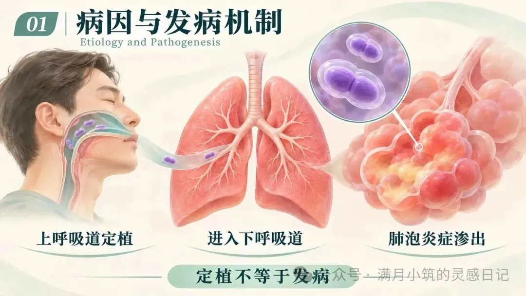
经典病理分为充血期、红色肝变期、灰色肝变期、消散期,各阶段没有清晰界限。早期规范使用抗菌药物后,完整典型分期很少见到。多数患者炎症吸收后肺组织可以完全复原,不留纤维化瘢痕;少数老年人吸收缓慢、吸收不全,可演变为机化性肺炎。大约5%~10%患者可并发脓胸,10%~20%患者细菌入血,引发心包炎、脑膜炎、关节炎等肺外感染。 Classic pathological changes include congestion phase, red hepatization phase, gray hepatization phase and resolution phase, without clear boundaries between stages. Complete typical staging is rarely seen after early standardized antibiotic therapy. Lung tissue can recover fully in most patients without fibrotic scars. A small number of elderly patients have slow or incomplete absorption and may develop organizing pneumonia. About 5%–10% of patients develop empyema, and 10%–20% develop bacteremia with extrapulmonary complications such as pericarditis, meningitis and arthritis. 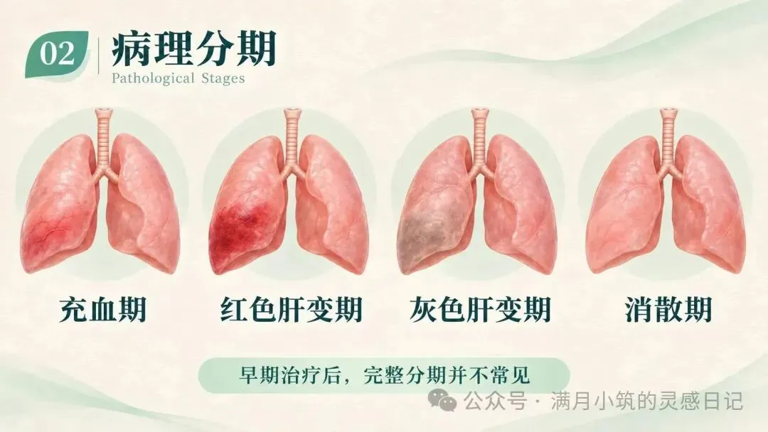
好发于冬春季节,青壮年、老年人、婴幼儿多见;吸烟、慢阻肺、心衰、免疫抑制人群属于高危人群。发病前常有受凉、劳累、醉酒、上感前驱史。典型起病急骤:高热寒战、肌肉酸痛、患侧胸痛,咳嗽伴铁锈色痰。体征有急性热病容、肺实变体征;重症可出现感染性休克、ARDS、意识改变。自然病程约1~2周,有效抗生素使用后体温常在1~3天内快速回落。 It occurs mostly in winter and spring, affecting young adults, the elderly and infants. Smokers, patients with chronic obstructive pulmonary disease, heart failure and immunocompromised individuals are high-risk groups. Preceding triggers include cold exposure, fatigue, alcohol intoxication or upper respiratory tract infection. Classic onset is abrupt: high fever, chills, myalgia, pleuritic chest pain and cough with rusty sputum. Signs include acute toxic appearance and consolidation signs. Severe cases may develop septic shock, ARDS and altered mental status. The natural course lasts about 1–2 weeks; fever usually resolves rapidly within 1–3 days after effective antibiotic administration. 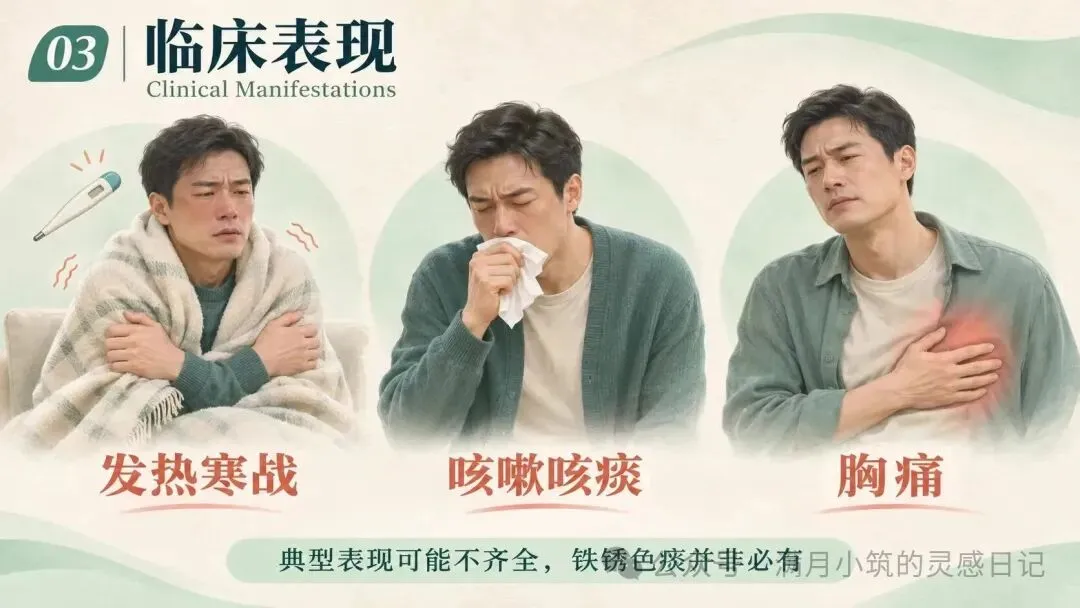
血常规提示白细胞升高、中性粒细胞占比高伴核左移;老年、重症、免疫低下患者白细胞可不升高,但中性比例仍然上升。痰革兰染色、荚膜染色、痰培养、PCR、抗原检测可以协助病原学确诊。胸片早期仅肺纹理增粗;进展后出现肺段、肺叶实变阴影,实变内可见支气管充气征,消散期可出现“假空洞征”。诊断结合症状体征+影像学,病原学阳性是确诊金标准;年老体弱、继发于其他疾病者表现常不典型,需要仔细鉴别。 Blood tests show elevated white blood cells, predominance of neutrophils with left shift. White blood cell count may be normal in elderly, severe or immunocompromised patients, while neutrophil percentage remains high. Sputum Gram stain, capsular stain, sputum culture, PCR and antigen assay help confirm the pathogen. Chest radiography only shows increased lung markings in early stage. Consolidation shadow limited to a lung segment or lobe appears as disease progresses, with air bronchogram inside consolidation. “Pseudocavity sign” can be seen during resolution. Diagnosis relies on clinical features plus imaging, and positive pathogen test is the gold standard for confirmation. Presentations are often atypical in frail elderly patients or those with underlying diseases, requiring careful differential diagnosis. 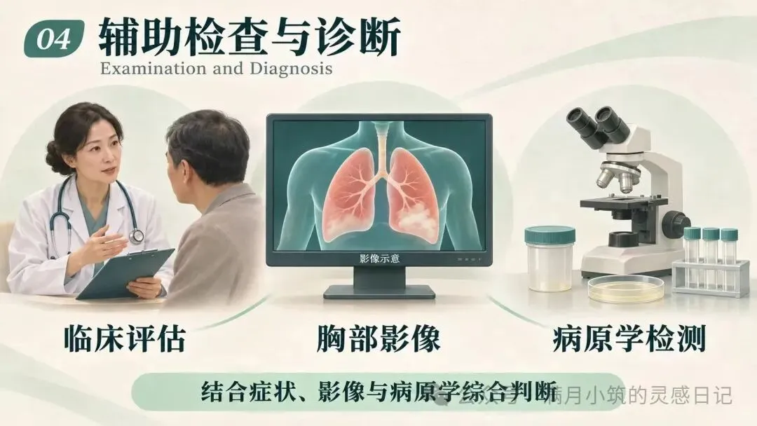
1. 抗菌治疗:首选青霉素,根据病情轻重选择剂型与剂量。青霉素耐药菌株、过敏患者,选用呼吸喹诺酮类、头孢曲松等;多重耐药菌株可选用万古霉素等。 Antibiotic therapy: Penicillin is the first-line agent. Formulation and dosage are adjusted according to disease severity. Respiratory fluoroquinolones or ceftriaxone are used for penicillin-resistant strains or penicillin-allergic patients. Vancomycin can be used for multidrug-resistant isolates. 2. 支持治疗:卧床休息,补充热量、蛋白、维生素;适当补液、氧疗,胸痛可酌情镇痛;尽量避免阿司匹林,防止干扰热型判断。镇静药物慎用,防止抑制呼吸。 Supportive care: Bed rest, adequate caloric, protein and vitamin supply. Fluid resuscitation and oxygen therapy when indicated. Analgesics may be used for chest pain. Aspirin should generally be avoided to prevent misinterpretation of fever curve. Sedatives are used cautiously to avoid respiratory depression. 3. 并发症处理:合并脓胸、胸腔积液,积极引流、送检培养;持续发热要警惕肺外感染或者其他合并病因。 Management of complications: Active drainage and specimen culture are required for empyema or pleural effusion. Persistent fever raises suspicion of extrapulmonary infection or alternative underlying causes. 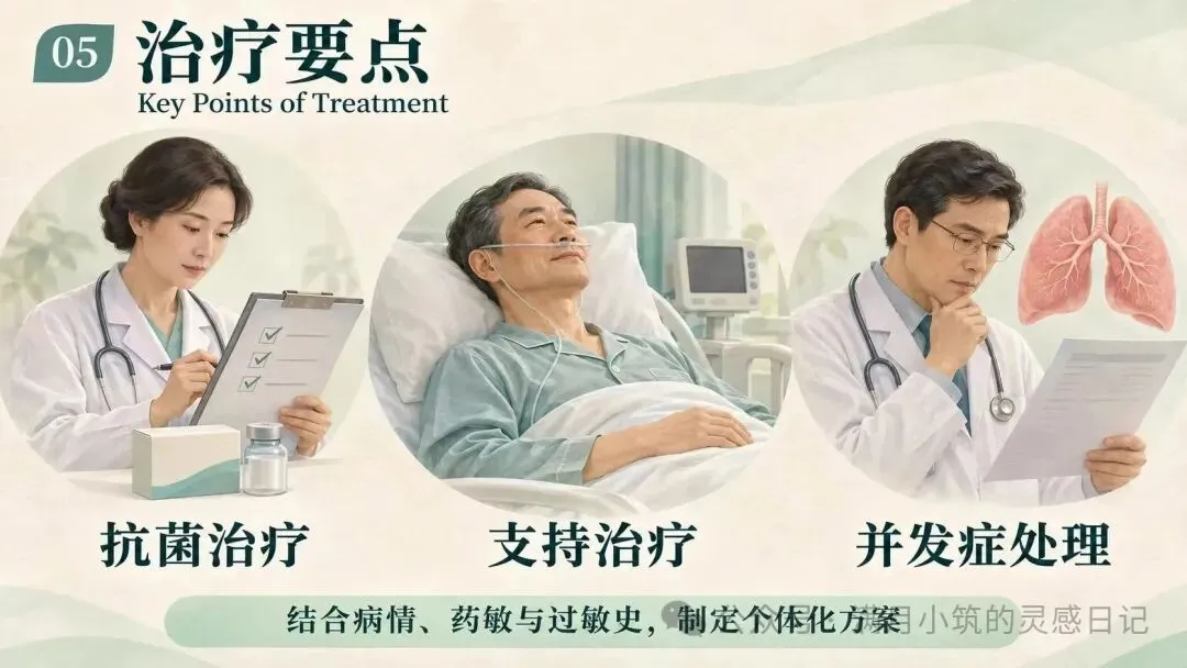
肺炎链球菌肺炎是CAP经典代表,核心考点:荚膜为毒力核心、不形成空洞、铁锈色痰、肺叶段实变、青霉素首选。现在典型病例越来越少,临床思维上不能只盯着经典表现,对于发热伴呼吸道症状患者,都需要把它纳入鉴别,尽早启动规范抗感染,警惕休克、脓胸等危重并发症。 Pneumococcal pneumonia is the classic representative of CAP. Core takeaways: capsule is the key virulence factor, no cavitation, rusty sputum, lobar consolidation, penicillin as first-line therapy. Classic cases are becoming less frequent. Clinicians should not only rely on typical manifestations. It should be included in differential diagnosis for patients with fever and respiratory symptoms. Standard anti-infection treatment should be initiated early, with vigilance for life-threatening complications such as shock and empyema.
一、病因与发病机制
Etiology and Pathogenesis
二、病理分期
Pathological Stages
三、临床表现
Clinical Manifestations
四、辅助检查与诊断
Auxiliary Examination and Diagnosis
五、治疗要点
Key Points of Treatment
六、小结
Summary
本文来自网友投稿或网络内容,如有侵犯您的权益请联系我们删除,联系邮箱:wyl860211@qq.com 。

